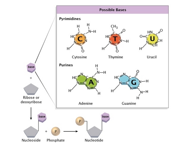
DNA is the molecule that stores the genetic instructions for all living organisms. The structure is a double helix made up of two long strands and four nitrogenous bases–guanine, cytosine, adenine, and thymine.
In 1953, Watson and Crick discovered the structure of DNA by building a physical model of its components. They were able to do this by analyzing the data gathered by Rosalind Franklin and Maurice Wilkins using X-ray diffraction techniques.
The Origins of the Double-Helix Model
The double helix model of DNA was a key discovery that revolutionized our understanding of genetics. It has helped explain how DNA replicates itself, transmits genetic information from one generation to the next, and performs all life’s essential functions.
The DNA molecule has two complementary strands that wind around each other in a right-handed double helix or a twisted ladder. Each strand has a sugar-phosphate backbone and nitrogenous bases (adenine, thymine, guanine, and cytosine) form base pairs that hold the two strands together.
Each rung of the ladder is made of two types of nitrogen bases, and each base pair is specific: adenine bonds to thymine, and guanine bonds to cytosine. These base pairings are a vital part of how the strands of DNA connect to each other, and they provide the structure for the digital code that tells the molecule how to replicate itself.
Maurice Wilkins, a crystallographer at King’s College London, and Rosalind Franklin were working with X-ray images of DNA. They were able to see that the strands were twisted, and they realized that the strands could only be held together by hydrogen bonds between the nitrogenous bases on each strand.
As a result, they proposed that DNA consists of two complementary strands twisted around each other to form a right-handed double helix. They also proposed that the helix comprises antiparallel 3′ – 5′ strands and that hydrogen bonds are formed between the nitrogenous bases on each strand.
This model was later credited to James Watson and Francis Crick, who worked on it in 1953. It was an important breakthrough in the field of biology and has influenced major advances in modern science, including gene mapping and gene therapy.
GEN’s John Sterling and Julianna LeMieux spoke to Matthew Cobb, professor of zoology at the University of Manchester, about the origins of the double helix model and how Rosalind Franklin played a critical role in the process.
The double helix has become one of biology’s most well-known and iconic structures. It has shaped many important developments in our understanding of genetics and has become an emblem for the sciences and the arts, often represented in statues, artwork, and toys.
Maurice Wilkins
Maurice Wilkins, born in Pongaroa, New Zealand, in 1916, was a British biophysicist who played a key role in the discovery of the structure of DNA. He shared the 1962 Nobel Prize in Physiology or Medicine with James Watson and Francis Crick for their contributions to understanding the molecular structure of deoxyribonucleic acid (DNA).
In 1950 Wilkins, then an assistant to John Randall at King’s College London began studying DNA fibers using X-ray diffraction techniques. He obtained high-resolution X-ray images of DNA fibers, and they suggested a helical corkscrew-like shape.
At the same time, Linus Pauling, the world’s leading physical chemist, discovered that proteins had single-stranded alpha helix structures, causing many biologists to think of DNA similarly.
Nevertheless, Franklin had made no progress in her work on protein, and in late 1950 she was asked by Wilkins to study X-ray diffraction data from the DNA fibers that Signer had sent her.
Although she had been working on the structure of proteins, Franklin was adamant that she should focus only on DNA, which seemed to her more interesting than the proteins. She had a theory that the sugar-phosphate backbones should be on the outside of the molecule, so she began to make a series of DNA experiments in which she tried to prove this.
But she soon became frustrated with her inability to find any progress on the fibers of DNA, and she began to suspect that she had been misled by her boss, Wilkins. She was wrong, but her misunderstanding led to an irrevocable breakdown in their working relationship and a serious breakdown of their friendship.
After his marriage to Franklin in 1951, Wilkins continued to research DNA at Kings College, and he became more interested in the structure of DNA as a molecule. He began to see it as the master molecule of life, and he decided that the structure of DNA must be simple enough to understand.
He was a pioneer in biophysics and was responsible for founding the MRC Cell Biophysics Unit at King’s College London. In later years, he devoted himself to the social responsibility of science and campaigned for nuclear disarmament.
Rosalind Franklin
Rosalind Franklin, who died in 1958, may not be as recognizable as James Watson and Francis Crick, but she was a crucial part of the discovery of DNA’s double-helix structure. Without her work, both of these scientists would have been unable to achieve their breakthroughs.
Born in London, England, Rosalind Franklin was a student of W.C. Price, a spectroscopist at Cambridge, where she studied within the Natural Sciences Tripos. She was also a member of the Natural History Society. She had a distinguished career in both coal chemistry and DNA research, but her most important scientific achievement was helping to uncover the double-helix structure of DNA.
In 1951, she took X-ray diffraction photographs of DNA, which J. D. Bernal called ‘amongst the most beautiful X-ray photographs of any substance ever taken’ and helped to show that there were two forms of DNA – wet and dry – with different structures. This led to her first major discovery – that there was another type of DNA, known as B-DNA, which sits in living cells exposed to high moisture levels.
Her X-ray images of the B form of DNA, known as an asymmetrical image, were very clear and helped to confirm the helical nature of DNA. Her asymmetrical image also showed that the four bases (adenine, thymine, guanine, and cytosine) were located on the outside of the DNA helix rather than the inside.
She continued to take pictures of the B form of DNA until she completed a series of experiments in 1952. The results of her experiments were shown to James Watson at King’s College, and the data confirmed the 3-D structure that Crick and Watson had theorized for DNA.
However, the relationship between Franklin and Wilkins was strained at this time. They did not communicate as well as they should have, and when Wilkins went on vacation, he assumed she was his assistant. When he returned, his mistrust of Franklin grew.
Despite the tensions between the two, Franklin stayed committed to her work on DNA. Her x-ray diffraction images were instrumental in the formation of the structure of DNA, and her data helped to drive Watson and Crick toward their discoveries.
James Watson and Francis Crick
In 1951, when a Canadian bacteriologist named Oswald Avery showed that DNA was the master molecule of life, James Watson was convinced that he had to figure out the structure of this molecule. He devoted his spare time to reading about genetics and molecular biology and talking with his friend, Nobelist Sydney Brenner.
At the same time, Rosalind Franklin and Maurice Wilkins were using X-rays to study DNA at King’s College London. They were able to shine X-ray beams through DNA crystals and record the resulting diffraction patterns on photographic film. They shared this data with Crick and Watson, who were both working at Cambridge University.
After seeing the X-ray images, Watson and Crick immediately realized that they had found a way to understand the structure of DNA. They had never studied DNA before, but their common physics and molecular biology backgrounds made them the perfect partners to pursue this goal.
The first step in their discovery was to build a model of DNA’s structure out of brass plates, clamps, and other bits of laboratory equipment. This was a huge breakthrough because it was the first time that they had seen the physical structure of DNA.
It was also the first time they had figured out the exact position of the two sugar-phosphate backbones of DNA. This made it possible to build a double helix model, which they published in Nature in 1953.
They based the model on the X-ray images and the chemistry of the DNA helix, but they needed to make it a physical model that could be tested for accuracy. They did this through experiments, but the most important part was that they could not have done it without each other.
In their experiment, they used the chemical bonds that formed between the hydrogen atoms of the bases in the DNA to determine the shape of the helix. This was the only way to prove that the helix was twisted into a double helix rather than a single helix.
Their discovery was a major step forward in understanding how DNA worked, and it opened the door for the rapid development of molecular biology that continues today. It also allowed scientists to understand how DNA was a key ingredient in hereditary information.